Blepharoplasty for Bilateral Upper Eyelids
Abstract
Upper blepharoplasty is one of the most commonly performed oculoplastic procedures. It is aimed at correcting the involutional changes of the upper eyelids, characterized by loose, excess eyelid skin (dermatochalasis) and preaponeurotic fat herniation (steatoblepharon) as well as some cases of ptosis. These conditions could result in functional symptoms, such as reduced visual fields, as well as cosmetic concerns and perceived body dysmorphia. In this case, the patient underwent upper blepharoplasty for cosmetic improvement and to remove xanthomatous lesions. This article discusses and demonstrates the preoperative assessment of the patient, the preparation, the surgical technique, and possible complications.
Case overview
Focused History of the Patient
Patients with dermatochalasis often report sagging or drooping of the eyelids, tired or sleepy appearance, obstruction of visual fields, brow-ache, and heavy sensation around the eyes.1,2 These symptoms can affect a patient's quality of life and self-confidence, and may lead them to seek medical or surgical intervention.3,4 Since the incentive behind the procedure is often cosmetic improvement, preoperative discussion of the expected results is imperative.3 Previous history of cosmetic procedures and the evaluation of patient’s perception of their outcomes could reveal unrealistic expectations.4,5 A thorough discussion can determine if upper blepharoplasty is the appropriate treatment for a patient's specific concerns and goals.
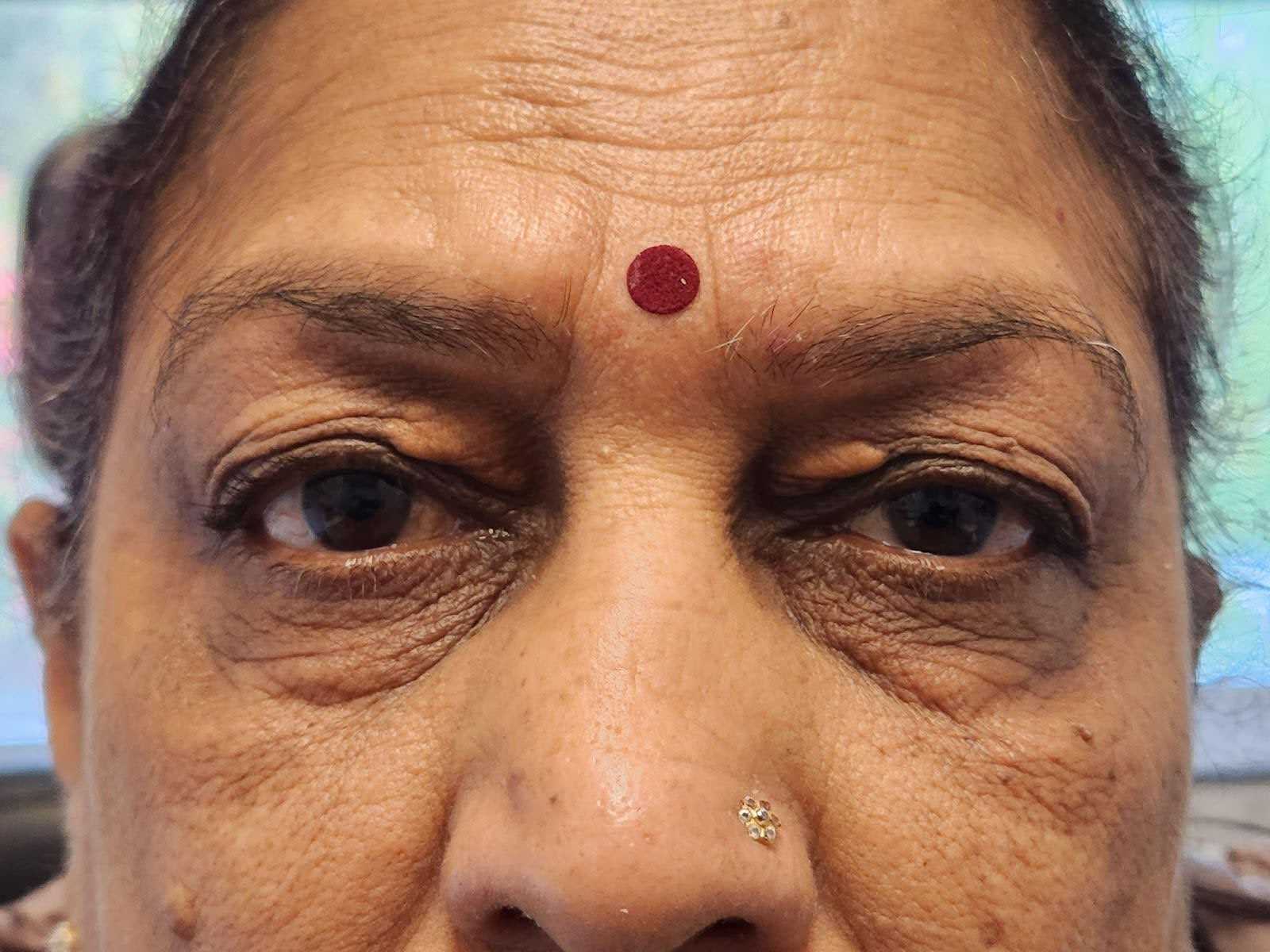
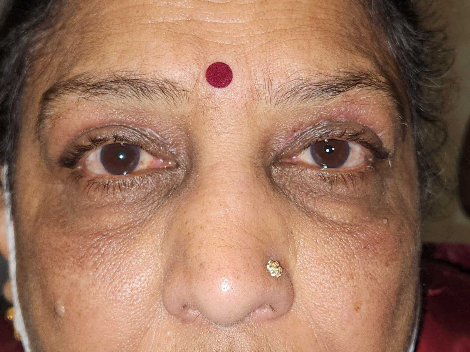
Figure 1. Patient before and after Surgery. Left) Patient Preoperatively. Note: xanthelasma in both eyes as well as the upper eyelid position. The upper eyelids are not crossing in front of the pupils, which is why this surgery was not covered by insurance and considered an elective procedure. It is possible to note dermatochalasis is unequal and worse in the left eye. Right) Patient 2 weeks postoperatively. Note: xanthelasma was successfully removed as well as improved upper eyelid position relative to the patients’ pupils.
Physical Examination
- Eyelid examination:
- Evaluation of eyelid position and their symmetry.
- Quality and quantity of redundant skin, evaluation of the preaponeurotic fat pads for the presence of herniation.
- Evaluation of the eye globe position in relation to the bony orbit in order to determine the optimal lid fold position.
- Evaluation for possible concurrent eyelid and brow ptosis.
- Lagophthalmos assessment, Bell’s phenomenon assessment.
- A complete ocular examination, including visual acuity testing, anterior segment examination, along with testing for dry eye disease, and fundus examination.
- Visual field testing with and without manual eyelid elevation.
- Preoperative photography.
- Medical history. A number systemic disease such as rheumatologic conditions, collagen vascular diseases, diabetes, and a history of keloids might lead to complications and inadequate wound healing.
- Blood thinning agents and other systemic medications should be noted.
Natural history
Dermatochalasis is a result of decreased skin elasticity due to age and gravity.6 Smoking and genetic predisposition also play a role.3 When left untreated it slowly progresses, gradually affecting the patient’s visual performance and quality of life.2
Options for treatment
Blepharoplasty is often considered the most effective and definitive treatment option for moderate to severe dermatochalasis. It addresses both the cosmetic and functional concerns associated with excess and sagging skin in the upper eyelids.3,6
Alternatives include non-invasive procedures such as radiofrequency,7 ultrasound, or laser therapy8 aimed at stimulating collagen production and improvement of skin texture and appearance. These treatments are however less effective for moderate or severe cases of dermatochalasis.
Topical pharmacological treatment of any ptosis component with Oxymetazoline hydrochloride ophthalmic solution, 0.1%. Mechanism of action is an alpha adrenergic agonist, which stimulates Mueller’s muscle and causes eyelid retraction.9
Rationale for Treatment
In this case blepharoplasty was performed in order to achieve cosmetic improvement and simultaneously remove xanthomatous lesions.
Special Considerations
- Blepharoplasty should not be performed in patients with uncontrolled systemic disease, which could lead to intra- and/or postoperative complications.
- Active eye infections should be treated before surgery.1
- Patients with a history of psychiatric disorders (e. g. body dysmorphic disorder, severe depression, obsessive-compulsive disorder etc.) or unrealistic expectations are bad candidates for blepharoplasty. History of dissatisfaction with previous cosmetic procedures can be another reason to delay or avoid surgery and opt for conservative treatments.3,4
Potential complications
Potential postoperative complications include the following:1,2
- Orbital hemorrhage and blindness
- Infection
- Bleeding
- Lagophthalmos, ocular surface exposure, and exposure keratopathy.
- Ptosis
- Ectropion or entropion
- Diplopia
- Numbness or decreased sensation
- Asymmetry
- Scarring
Discussion
The excess skin is marked prior to local anesthetic injection. The amount of skin to be removed is determined with a “pinch test”—the skin is held with toothless forceps and lifted to determine the amount of redundancy. Care should be taken not to remove too much skin, at least 20–24 mm of skin should be left between the inferior border of the brow and the upper lid margin.1,5 The medial extent of the incision margin marking should not extend further than the punctum, and the lateral end of the marking should be kept within the lateral orbital rim. After the incision site is marked, the patient is anesthetized with a local injection of lidocaine and epinephrine. After the patient is draped, an incision is made along the marked line with a Bard-Parker No. 15 blade, and the skin flap is removed with a blade or scissors. If necessary, fat pads can be removed at this stage. In this case, cholesterol plaques were removed. After cautery, the skin is closed with a running suture. Special techniques should be used when operating on patients of Asian descent. The incision is usually made closer to the lid margin and some preaponeurotic fat is to be preserved to maintain the Asian-type lid structure if the patient wishes.10,11
Equipment
- Surgical marker
- No.15 Bard-Parker blade
- Forceps
- Scissors
- Cautery
- 6-0 sutures
Disclosures
Nothing to disclose.
Statement of Consent
The patient referred to in this video article has given their informed consent to be filmed and is aware that information and images will be published online.
Citations
- Korn BS. 2021-2022 Basic and Clinical Science Course, Section 7: Oculofacial Plastic and Orbital Surgery. American Academy of Ophthalmology, Section 7. Published online 2021.
- Marco K, Andrea B, Andrea R, Valeriano V. Aesthetic Plastic Surgery. In: Textbook of Plastic and Reconstructive Surgery. Springer International Publishing; 2022:509-520. doi:10.1007/978-3-030-82335-1_33.
- Genç ÇD. Upper eyelid blepharoplasty results and evaluation of patient satisfaction. Eur J Pub Health Stud. 2022;5(2). doi:10.46827/ejphs.v5i2.129.
- Joseph AW, Ishii L, Joseph SS, et al. Prevalence of body dysmorphic disorder and surgeon diagnostic accuracy in facial plastic and oculoplastic surgery clinics. JAMA Facial Plast Surg. 2017;19(4). doi:10.1001/jamafacial.2016.1535.
- Rumelt S. Ophthalmology, 4th Edition, (in print and online) Yanoff M, Duker JS. (2013) ISBN 978-1455-7398-44, Elsevier. Graefe’s Archive for Clinical and Experimental Ophthalmology. 2017;255(2). doi:10.1007/s00417-015-3050-y.
- Raschke GF, Bader RD, Rieger UM, Schultze-Mosgau S. Photo-assisted analysis of blepharoplasty results. Ann Plast Surg. 2011;66(4):328-333. doi:10.1097/SAP.0b013e3181fadd71.
- Verner I, Naveh HP, Cotofana S. A novel ablative radiofrequency microplasma nonsurgical blepharoplasty for dermatochalasis. Dermatol Ther. 2020;33(6). doi:10.1111/dth.14002.
- Bae-Harboe YSC, Geronemus RG. Eyelid tightening by CO2 fractional laser, alternative to blepharoplasty. Derm Surg. 2014;40:S137-S141. doi:10.1097/DSS.0000000000000165.
- American Board of Cosmetic Surgery. Here’s what you need to know about Upneeq, the FDA approved “eyelid” lifting drops. Accessed 08/11/23. Available at: https://www.americanboardcosmeticsurgery.org/news/upneeq-fda-approved-eye-lifting-drops.
- Paik JS, Lee JH, Uppal S, Choi WC. Intricacies of upper blepharoplasty in Asian burden lids. Fac Plast Surg. 2020;36(5). doi:10.1055/s-0040-1718391.
- Chee E, Choo CT. Asian blepharoplasty - an overview. Orbit. 2011;30(1). doi:10.3109/01676830.2010.535644.


| Publication Date | 11/30/2023 |
| Article ID | 400 |
| Production ID | 0400 |
| Volume | 2023 |
| Issue | 400 |
| DOI | |
| https://doi.org/10.24296/jomi/400 | |