The Use of Photodynamic Nails for Bone Reinforcement in Combination with Complex Total Hip Arthroplasty in the Setting of Radiation Osteitis
Abstract
Herein, we present a case of diffuse large B-cell lymphoma (DLBCL) with skeletal involvement in a geriatric male. Initially presenting with left hip pain, the patient was diagnosed with DLBCL affecting the left acetabulum. Subsequent treatment with systemic and radiation therapy resulted in radiation osteitis, osteoarthritis, and acetabular collapse, necessitating surgical intervention.
The treatment plan involved total hip arthroplasty (THA) with photodynamic intramedullary nails (PDNs) for pelvic stabilization, augmented with tantalum augments for enhanced support. PDNs provided structural stability while minimizing interference with future oncological interventions. The surgical procedure comprised meticulous insertion of PDNs and placement of tantalum augments, achieving optimal stability and alignment of the acetabular component.
This case underscores the strategic use of PDNs and tantalum augments in for treating major acetabular defects in patients with complex pathologies who require THA for pelvic stabilization. These techniques provide advantages in postoperative radiographic disease monitoring and precision in radiation therapy planning. The multidisciplinary approach emphasizes the importance of carefully selecting the appropriate implants to optimize outcomes in orthopaedic oncology.
Keywords
Pelvic stabilization; photodynamic nails; radiation osteitis; complex total hip arthroplasty.
Case Overview
Background
Addressing diffuse large B-cell lymphoma (DLBCL) with skeletal involvement demands a nuanced approach, considering the interplay of disease progression, lesion location, patient characteristics, and treatment options. While the treatment paradigm has evolved towards non-operative management, encompassing advanced systemic chemotherapy and radiation therapy, the potential secondary effects of these therapies warrant careful consideration, particularly considering improved patient survival. Patients with acetabular lesions and/or radiation osteitis present unique challenges as older, comorbid patients with compromised bone integrity may be unsuitable for isolated THA. Therefore, less invasive strategies offering structural stability and restoration of biomechanics are highly desirable, such as percutaneous placement of photodynamic intramedullary nails for pelvic stabilization in isolation or before complex THA. Stabilization with photodynamic balloons does not preclude future arthroplasty or reconstruction.
Focused History of the Patient
A geriatric white male presented with a complaint of hip pain, which, upon biopsy, revealed DLBCL of the left acetabulum. He underwent systemic and radiation therapy for lymphoma, showing a favorable response. However, subsequent imaging indicated a loss of acetabular integrity and superior migration of the femoral head within the anterior column, attributed to radiation-induced osteitis. Following the completion of systemic and radiation therapy, surgery was planned to address the radiation osteitis and resultant leg length discrepancy. This involved complex THA with stabilization of the pelvic columns using PDNs. Percutaneous application of PDNs was utilized to augment the stability of the THA, incorporating tantalum augments for enhanced support.
Physical Exam
The musculoskeletal examination of the lower extremities six months before surgical intervention revealed a normal but mildly antalgic gait, particularly over long distances. After walking around 100 to 150 yards, the patient experienced bilateral hip pain, which was more severe in the left hip. Palpation revealed no edema or tenderness. Range of motion was restricted in both hips with decreased internal rotation of the left hip, measuring approximately 10 to 15 degrees, while external rotation remained preserved at 45 degrees bilaterally. There were no restrictions on hip flexion, knee flexion and extension, or ankle flexion and extension. Neurologically, the patient exhibited normal muscle strength and sensation across L1–S2 myotomes and dermatomes, respectively, with no deficits observed. Numbness at the bottom of the feet was attributed to the effect of chemotherapy (e.g., cyclophosphamide, doxorubicin, prednisone, rituximab, and vincristine). Vascular examination revealed palpable dorsalis pedis and posterior tibial pulses, while skin integrity was intact throughout the lower extremities.
Upon subsequent examination three months prior to surgery, notable reductions in flexion, internal rotation, external rotation, and abduction were observed in the left hip. Furthermore, significant limb shortening was attributed to cranial migration of the femoral head.
Imaging
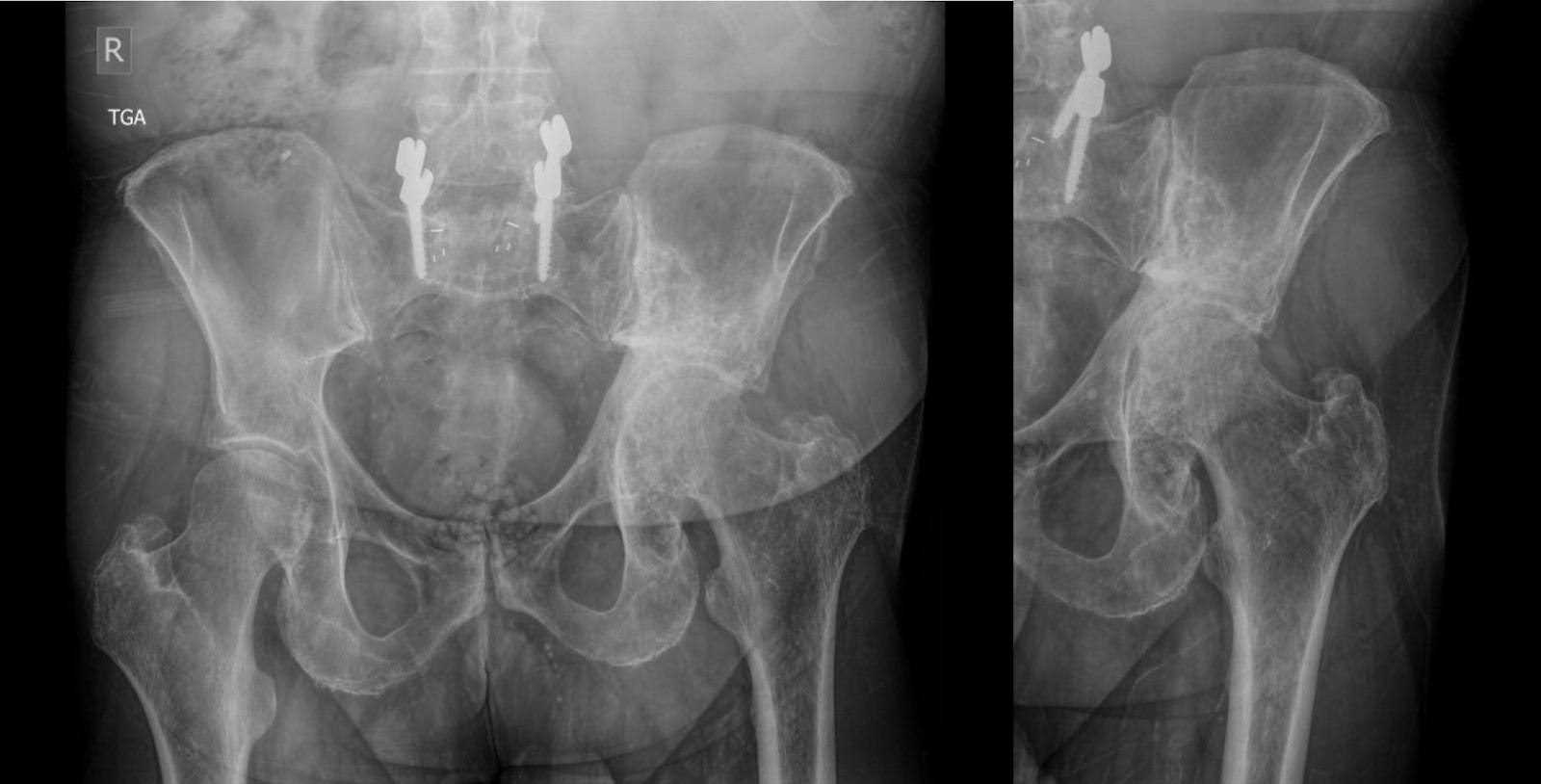
Upon presentation, X-ray imaging of the pelvis demonstrated left hip loss of joint space, osteophytes, subchondral cysts, and proximal migration of the femur with sacroiliac joint changes. Mild superior erosion of the acetabulum accompanied by sclerosis attributed to lymphoma. Degenerative changes in the sacroiliac joint and pubic symphysis were also present. Additionally, transpedicular fixation at the lumbosacral junction with interbody graft markers stemming from lumbar fusion surgery performed eight years earlier for lumbar degenerative disk disease.
Subsequent X-ray imaging, conducted three months preoperatively, revealed no substantive change in the appearance of mixed sclerotic and lytic lesions within the left hemipelvis, in keeping with treated lymphoma. This was accompanied by acetabular remodeling and cranial migration of the proximal femur, with degenerative changes in the left hip joint (Figure 1). Computed tomography (CT) findings aligned with those on X-ray, affirming the observed pathology (Figure 2).

Figure 1. Anteroposterior (AP) X-rays of the pelvis three months prior to surgery. Primarily sclerotic lesion of the left hemipelvis, consistent with treated lymphoma. Structural changes of the acetabulum with remodeling and superior migration of the proximal femur.

Figure 2. Axial, coronal, and sagittal views of CT of the pelvis three months prior to surgery. Mixed sclerotic/lytic lesion of the acetabulum redemonstrating treated lymphoma. Collapse of the acetabular roof and migration of the proximal femur into the supra-acetabular area with protrusion.
Natural History
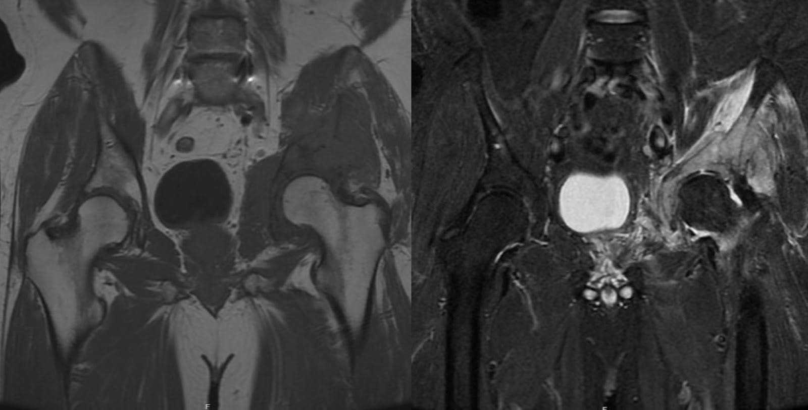
DLBCL is the most prevalent subtype of non-Hodgkin lymphoma, constituting approximately 30–40% of all cases.1 Diagnosis commonly occurs between the fifth and sixth decades of life. Its etiology is multifactorial and may involve genetic predispositions, immune dysregulation, as well as viral, environmental, and occupational exposures.2,3 It is marked by the proliferation of lymphopoietic cells, often originating within the bone, and has the potential to cause localized destruction of bony architecture, ultimately predisposing an individual to pathologic fractures.4 Clinically, DLBCL can present with skeletal pain secondary to bone destruction and destabilization. This pain may radiate, particularly when there is involvement of localized soft tissue components such as nerves, muscles, or vessels, depending on the lesion location. Systemic symptoms such as fever, night sweats, and weight loss can also be part of the constellation of symptoms.2,4,5 Imaging often reveals radiolucent bony destruction with soft tissue involvement, evident on plain film. Magnetic resonance imaging (MRI) typically shows relative hypotintensity on T1 sequences in the medullary canal, indicating bone marrow replacement. Additionally, T2 sequences commonly display hyperintensity within both intramedullary and extramedullary extension.6 Approximately four years before the pelvic stabilization surgery, the patient’s MRI revealed a T1 hypointense and T2 hyperintense lesion in the left acetabulum and innominate bone, consistent with a pathological process in these areas (Figure 3). The prominent soft tissue masses frequently observed at presentation for DLBCL can progress without systemic intervention. Standard treatment modalities for DLBCL involve chemotherapy and localized radiation therapy.7 DLBCL has a moderate to favorable prognosis, with 5-year survival rates ranging from 60–70% following first-line therapy.1

Figure 3. Preoperative MRI, taken approximately four years before pelvic stabilization surgery. MRI showed a T1 hypointense and T2 hyperintense lesion in the left acetabulum and innominate bone, suggestive of a pathological process.
Although lymphoma is generally responsive to radiation therapy, long-term effects on bone, known as radiation osteitis, remain incompletely understood. Radiation osteitis may manifest as osteopenia, disruption of trabecular architecture, and cortical irregularities, rendering the bone susceptible to fractures and abnormal remodeling. These alterations predispose the affected bone to fractures and aberrant remodeling processes. This risk intensifies in long bones and weight-bearing regions like the acetabulum, potentially culminating in progressive osteoarthritis.8,9 Additionally, radiation involving chondral cells substantially contributes to these degenerative alterations.10
THA remains a widely used and dependable surgical intervention for osteoarthritis, encompassing a spectrum of complexities from primary degenerative osteoarthritis to cases involving pelvic deformities and defects due to prior diseases or other pathology.11–13 Advances in metallurgy and material science have enhanced THA durability since its widespread adoption. Augments, a technique utilized to address pelvic defects, facilitate bony defect filling by integrating the hemispherical shell of the acetabular component with a trabecular metal face, promoting biological ingrowth, and securing with screw fixation.14,15 Augmentation methods, including cement and photodynamic balloons, offer structural support, which is particularly crucial for oncology patients. Photodynamic balloons inserted percutaneously provide stability for the acetabular cup, complementing the function of augments. This combination becomes increasingly significant as targeted carcinoma therapy improves and life expectancy rises, achieving pain relief and rectifying acetabular defects, as evidenced by addressing leg length discrepancy in specific cases.16,17
Options for Treatment
Multiple treatment options exist for hip osteoarthritis, ranging from steroid injections to surgical intervention.18 However, in cases characterized by multifocal pathologies, such as radiation osteitis and superior migration of the femoral head, joint reconstruction emerges as the optimal treatment option. Hemiarthroplasty is not a suitable option due to the non-hemispherical and eroded acetabulum. Although THA in situ is an option, it may worsen the limb length discrepancy and alter biomechanics, leading to increased instability and dislocation risk.19 Reconstruction with a jumbo acetabular component may address the joint space alteration and large defect, but it necessitates extended acetabular reaming, resulting in potential bone loss.20
Conversely, utilizing a standard-size acetabular component with augments offers bone preservation, though screw fixation may be hindered by radiation osteitis. Overcoming this challenge requires meticulous attention to achieving adequate screw depth. Other reconstructive strategies rely on large custom triflange constructs or complex cup cage constructs. These options are effective but have a significant risk of infection and instability.21,22 Cup cage constructs are also an option for large acetabular reconstruction but carry the risk of instability and infection.23 These large constructs can increase intraoperative morbidity and complicate radiographic disease monitoring after surgery.24–26 Utilizing PDNs as augments is a minimally invasive alternative that can provide fixation connected to an endosteal strut spanning the pelvic column, effectively restoring the center of rotation.27
Rationale for Treatment
In cases of pelvic lymphoma, the bony architecture may collapse during the time it takes for radiation and chemotherapy to take effect, particularly in weight-bearing joints. As any pressure is exerted into the structurally weakened acetabulum, the femoral head may migrate proximally upward, causing limb length discrepancy and a restricted range of motion. Systemic therapies, however, allow for bony healing with disease treatment and osseous consolidation. Radiation osteitis, combined with limited range of motion, can speed acetabular wear, leading to pain. The priority of THA is pain alleviation, followed by correction of the limb length discrepancy. This approach may be further reinforced by PDN stabilization of the acetabulum, thereby enhancing treatment outcomes and improving patient quality of life.
Special Considerations
PDNs offer a versatile solution for acetabular reconstruction, serving as primary stabilizers of the pelvic columns while facilitating secure fixation of implants in reconstructive procedures. Their exceptional resistance to compressive, torsional, and tensile forces and the ease of delivery via a flexible catheter enable precise anatomic restoration of the acetabular column. With the flexible insertion and curing of the PDN after volumetric filling, there are multiple points of contact within bone, leading to overall improved stability and lower risk of mechanical failure due to stress concentration within the implant.28 Additionally, their radiolucency permits clear imaging without metal artifact interference during radiographic disease monitoring. PDNs allow for the fixation of screws within the cured material, facilitating seamless integration with endoprosthetic constructs while preserving the potential for local osseointegration.16 Their superior longitudinal strength and rotational stability eliminate the need for additional screw stabilization and effectively distribute mechanical resistance throughout the implant. Additionally, the mechanical characteristics of PDNs are closer to bone compared with metal and thus have a lower risk of stress shielding, leading to a better integrated construct within bone.28 Despite these advantages, the encasement of PDNs within a polyethylene balloon catheter may restrict bony ingrowth. However, the absence of cement or similar substrates may promote greater osseointegration compared to conventional constructs.16
Surgical Procedure
This complex hip replacement involved reconstructing the joint, reinforcing the pelvis with PDNs, and neurolyzing the sciatic nerve. The procedure was performed under general anesthesia, and the patient was classified as American Society of Anesthesiologists (ASA) Physical Status III.
The patient was initially positioned prone, with compression boots applied bilaterally. All bony prominences were appropriately padded for protection. Secure fixation was ensured with chest rolls on a flat Jackson table. Preoperative prophylactic antibiotics (2 g of Ancef) were administered, with subsequent redosing every four hours throughout the procedure.
A small transverse incision was carefully made at the right posterior inferior iliac spine, followed by the placement of a navigation tracker and acquisition of an intraoperative O-arm spin. Utilizing navigation guidance alongside fluoroscopy, additional incisions were made at the left posterior inferior iliac spine and in the ischial prominence. Subsequently, a 3.2-mm drill bit was meticulously advanced to delineate the trajectories of the balloons. Employing a straight awl, we ensured precise positioning over the entry point while the 3.2-mm drill bit served as a guide. Upon confirmation of optimal wire placement in the supra-acetabular area and posterior column via fluoroscopic imaging in several views including iliac oblique, anterior-posterior pelvic, inlet, and obturator oblique, the drill bit was exchanged for a 2-mm guidewire. Following this, trajectory reaming for both balloons was conducted with careful attention to detail.
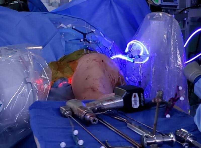
Debridement posed considerable challenges owing to sclerosis and the patient's rapid postradiation osteitis progression. Two balloons were sized: one measuring 22 mm x 140 mm for the supra-acetabular area and another sized 22 mm x 120 mm for the posterior column. Balloon insertion proceeded, followed by inflation with polymer, ensuring optimal filling of trajectories and bone defect regions. Polymer curing proceeded without complications (Figure 4). The placement system was subsequently excised for both balloons, with thorough irrigation of both sites. Closure involved layered suturing, utilizing 0 polydioxanone (PDS) for deep layers and 2-0 PDS for superficial layers. Skin closure was achieved using 3-0 Monocryl, Dermabond, Telfa, and Tegaderms for optimal wound management.

Figure 4. Curing Process. PDNs light annealing with fluoroscopy in place to monitor implant inflation. (Repurposed with permission from Fourman MS, Ramsey DC, Newman ET, Raskin KA, Tobert DG, Lozano-Calderon S. How I do it: percutaneous stabilization of symptomatic sacral and periacetabular metastatic lesions with photodynamic nails. J Surg Oncol. 2021;124(7):1192-1199. doi:10.1002/jso.26617.).
At this point, the patient was transitioned to lateral decubitus position using a hip grip on the same flat Jackson table. A longitudinal incision following the posterolateral approach to the left hip was made with a 10-blade, and subsequent dissection of subcutaneous tissues was carried out using electrocautery. The fascia was incised longitudinally, and the gluteus maximus was split, with detachment of the upper 50% of the sling and the external rotators as a single unit with the capsule. Given significant pelvis overgrowth, an in situ cut was made after placing two Cobra retractors, followed by identification of the intramedullary portion of the bone using a canal finder.
Sequential broaching of the femur up to size 6 stem was performed. This resulted in excellent restoration of anteversion and a satisfactory feeling of the canal, with no discernible mobilization of the trial upon internal and external rotation. The broach was left in situ to minimize bleeding.
The posterior column was fully exposed, and the sciatic nerve was identified for neurolysis from the sciatic notch down to the proximal thigh, anticipating the lengthening expected during reconstruction. Upon head removal, complete visualization of the acetabulum, including its superior center of migration, was achieved. Sequential reamers were utilized, reaming at the lowest point of the acetabulum with the transverse ligament serving as an anatomical reference for anteversion and abduction determination. Sequential reaming began at 44 mm to medially realign the native acetabulum, progressing up to size 54 mm. An augment measuring 15 mm in thickness was secured using three 6.5-mm screws measuring 30, 45, and 40 mm in length, achieving excellent fixation.
The acetabular surface was prepared until bony bleeding was achieved. Subsequently, the acetabulum was packed with 30 cc of cortical cancellous bone graft. Following this, a 56 multihole revision cup was inserted and secured with eight 6.5-mm screws ranging in length from 15 to 50 mm in diameter. The compatibility of a dual mobility cup with a -4 dual mobility and 28/52-mm stem was determined suitable for fixing the limb length discrepancy. The final components were smoothly inserted without difficulty, with intraoperative X-rays confirming proper implant positioning. Copious irrigation was performed, and no drains were used. Satisfactory hemostasis was achieved, and repair of the external rotators and capsule was performed using bone tunnels and #5 Ethibond stitches. Closure of the interval between the external rotators and the gluteus minimus was accomplished with interrupted #1 PDS stitches. Subsequently, #1 PDS was used for deep fascial layers. The deep subcutaneous layer was closed with 0 PDS interrupted stitches, and the superficial layer with 2-0 PDS interrupted stitches. Skin closure was completed with 3-0 Monocryl and Dermabond, followed by the application of a sterile dressing with Telfa and Tegaderm. The patient emerged from anesthesia without complications, maintaining neurovascular integrity and restoration of left lower extremity length. The patient exhibited a hip flexion contracture necessitating physical therapy; however, a small release of the anterior capsule proved insufficient given the contracture severity. The case length was 386 minutes, with an estimated blood loss of 400 mL. The patient remains alive 18 months postoperatively, with formal follow-ups at two weeks, six weeks, three months, four months, six months, and nine months. At the latest follow-up, he reported improved pain control, regained ambulation, walked his dog daily, and completed over 30 physical therapy sessions.
Discussion
Here we present the case of a geriatric male with left acetabular DLBCL. The patient responded favorably to chemotherapy and radiation therapy; however, subsequent follow up revealed persistent collapse of the acetabulum and superior migration of the femoral head. These changes were attributed to radiation osteitis following treatment and collapse from disease prior to full treatment effect. To address the resulting leg length discrepancy, biomechanical disruptions, and pain, the patient underwent percutaneous stabilization with PDNs to enhance the fixation quality of a complex THA performed utilizing tantalum augments.
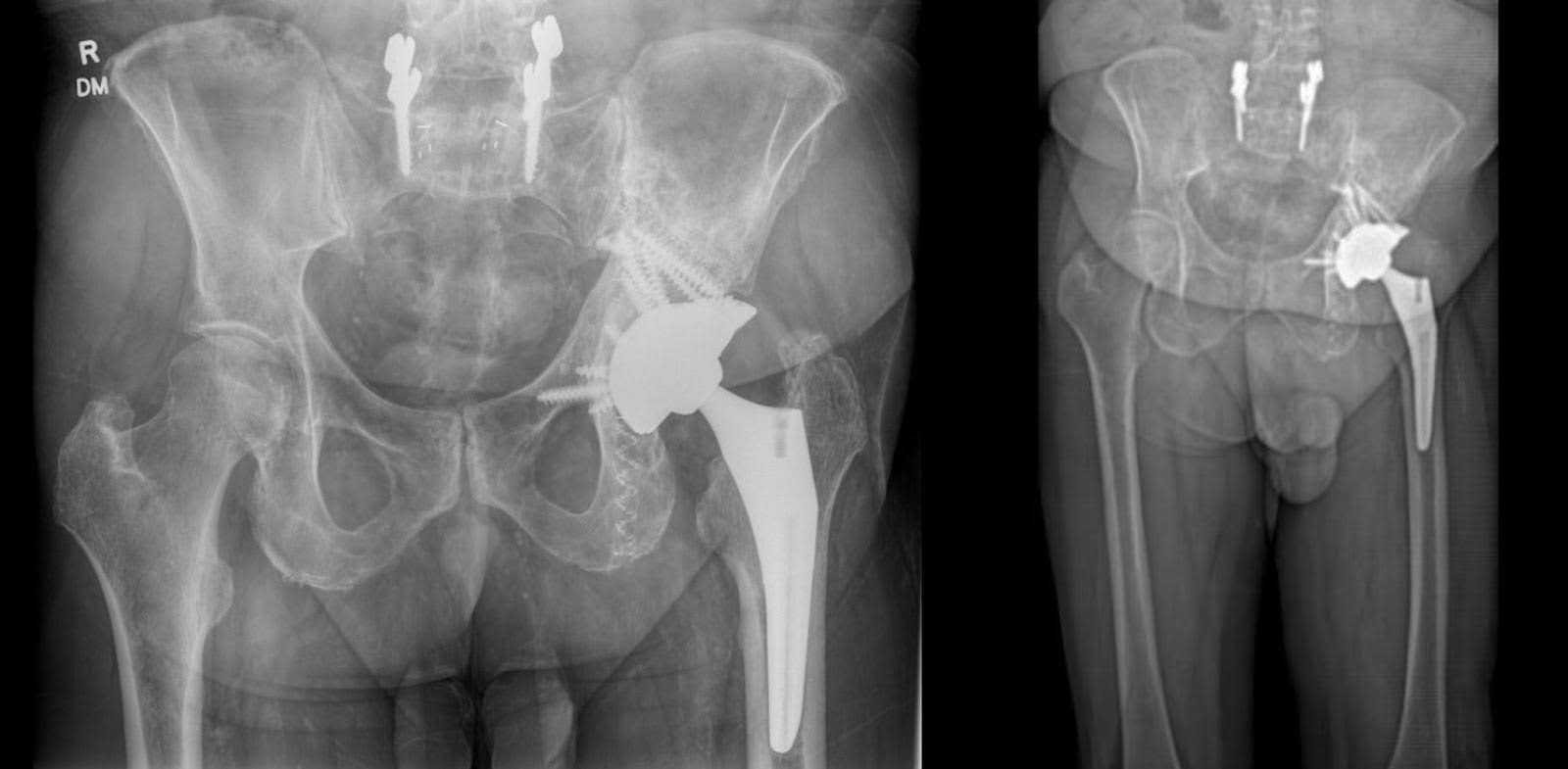
Six months after surgery, the patient demonstrated a full range of motion in the left hip, albeit reporting localized pain during internal rotation. Hip flexion and extension were within normal limits. Clinically, the patient exhibited nearly appropriate leg length, yet displayed an antalgic gait with reduced left stride length compared to the right. Despite participating in over 30 physical therapy sessions, the patient experienced moderate fatigue and pain during ambulation, necessitating frequent breaks after walking moderate distances. Left groin and lateral hip pain still increases during translational and prolonged periods of activity. X-rays of the hip joint and pelvis revealed a well-aligned left THA with tantalum augments in optimal positions, showing no signs of loosening (Figure 5).

Figure 5. AP X-ray of the pelvis six months after surgery. Stable alignment following left total hip arthroplasty with acetabular augmentation, alongside fixation of the left ischial and iliac bone using photodynamic nails. No evidence of periprosthetic fracture.
Lymphoma with skeletal involvement imposes a multifaceted challenge on orthopaedic surgeons, extending their responsibilities beyond primary tumor management to address the late effects of systemic chemotherapy or radiation therapy. This disease necessitates a comprehensive, multidisciplinary approach that integrates the expertise of orthopaedic surgeons, medical oncologists, and radiation oncologists. Central to this approach is the utilization of well-established immunohistochemistry markers, such as CD20 expression, which not only assist in diagnosing DLBCL but also inform the selection of appropriate treatment regimens, namely the widely employed R-CHOP chemotherapy protocol.29 While these treatments are crucial for disease control, they can significantly impact bone structure and function. Radiation therapy, for instance, may shrink lesions by directly killing lymphoma cells or disrupting their genetic material. However, it can also alter the primary structure of bone collagen, degrade cartilage, and induce radiation osteitis.9,30 Consequently, the weakened or loss of bone stock may predispose patients to subsequent osteoarthritis, often necessitating joint reconstruction through arthroplasty.
THA is an effective and well-tolerated surgery for treating osteoarthritis. However, it can be a technical challenge to perform when acetabular defects are present. There are various techniques for reconstructing acetabular defects, each with its own advantages and disadvantages.31 Therefore, there is no single best option for addressing acetabular defects, especially in oncology patients. Various techniques are published, including cup cage constructs, custom implants, and augment applications.21,23,32 A promising technique for reconstructing acetabular defects involves utilizing PDNs to reconstruct the acetabular architecture and using it as a scaffold for internally fixing the acetabular component.
Future advancements in THA and acetabular reconstruction offer promising prospects with the emergence of new technologies addressing bone defects in acetabular components. The evolution of custom implants utilizing 3D printing technology represents a significant avenue toward enhancing the efficiency and expediency of total hip revision arthroplasty. Furthermore, the utilization of bone substitutes in THA is increasingly viable as advancements in bone substitute materials continue to progress.33 Additionally, the amalgamation of metal mesh with impaction bone grafting has been delineated as an alternative approach, showcasing promising outcomes with mid to long-term follow-up.34
Equipment
Specialized equipment required for the procedure includes photodynamic balloons and the accompanying monomer for injection. Moreover, a light source unit is essential for the curing process of the PDN. A radiolucent table is indispensable for PDN insertion, particularly as pelvic utilization necessitates radiographic visualization, facilitated by either fluoroscopy or intraoperative CT scan for navigation. The preference of the author leans towards intraoperative CT navigation due to its ability to enhance drilling accuracy, especially in cases where compromised bone stock impairs tactile feedback.
Disclosures
The corresponding author (SALC) receives research support from and serves as a paid speaker and consultant for IlluminOss Medical Inc.
Statement of Consent
The patient referred to in this video article has given their informed consent to be filmed and is aware that information and images will be published online.
Citations
- Li S, Young KH, Medeiros LJ. Diffuse large B-cell lymphoma. Pathology. 2018;50(1):74-87. doi:10.1016/j.pathol.2017.09.006.
- Sehn LH, Salles G. Diffuse large B-cell lymphoma. N Engl J Med. 2021;384(9):842-858. doi:10.1056/NEJMra2027612.
- Cerhan JR, Kricker A, Paltiel O, et al. Medical history, lifestyle, family history, and occupational risk factors for diffuse large B-cell lymphoma: the InterLymph Non-Hodgkin Lymphoma Subtypes Project. J Natl Cancer Inst Monogr. 2014;2014(48):15-25. doi:10.1093/jncimonographs/lgu010.
- Yohannan B, Rios A. Primary diffuse large B-cell lymphoma of the bone. J Hematol. 2023;12(2):75-81. doi:10.14740/jh1087.
- Baar J, Burkes RL, Gospodarowicz M. Primary non-Hodgkin’s lymphoma of bone. Semin Oncol. 1999;26(3):270-275.
- Shah HJ, Keraliya AR, Jagannathan JP, Tirumani SH, Lele VR, DiPiro PJ. Diffuse large B-cell lymphoma in the era of precision oncology: how imaging is helpful. Korean J Radiol. 2017;18(1):54-70. doi:10.3348/kjr.2017.18.1.54.
- Susanibar-Adaniya S, Barta SK. 2021 update on diffuse large B cell lymphoma: a review of current data and potential applications on risk stratification and management. Am J Hematol. 2021;96(5):617-629. doi:10.1002/ajh.26151.
- Fu AL, Greven KM, Maruyama Y. Radiation osteitis and insufficiency fractures after pelvic irradiation for gynecologic malignancies. Am J Clin Oncol. 1994;17(3):248-254. doi:10.1097/00000421-199406000-00015.
- Willey JS, Lloyd SAJ, Nelson GA, Bateman TA. Ionizing radiation and bone loss: space exploration and clinical therapy applications. Clin Rev Bone Miner Metab. 2011;9(1):54-62. doi:10.1007/s12018-011-9092-8.
- Margulies BS, Horton JA, Wang Y, Damron TA, Allen MJ. Effects of radiation therapy on chondrocytes in vitro. Calcif Tissue Int. 2006;78(5):302-313. doi:10.1007/s00223-005-0135-3.
- Ferguson RJ, Palmer AJ, Taylor A, Porter ML, Malchau H, Glyn-Jones S. Hip replacement. Lancet. 2018;392(10158):1662-1671. doi:10.1016/S0140-6736(18)31777-X.
- Stiehl JB, Saluja R, Diener T. Reconstruction of major column defects and pelvic discontinuity in revision total hip arthroplasty. J Arthroplasty. 2000;15(7):849-857. doi:10.1054/arth.2000.9320.
- Walker RH. Pelvic reconstruction/total hip arthroplasty for metastatic acetabular insufficiency. Clin Orthop Relat Res. 1993;(294):170-175.
- Melnic CM, Salimy MS, Hosseinzadeh S, et al. Trabecular metal augments in severe malignancy-associated acetabular bone loss. Hip Int. 2023;33(4):678-684. doi:10.1177/11207000221110787.
- Karuppal R. Biological fixation of total hip arthroplasty: facts and factors. J Orthop. 2016;13(3):190-192. doi:10.1016/j.jor.2016.06.002.
- Heng M, Fourman MS, Mitrevski A, Berner E, Lozano-Calderon SA. Augmenting pathologic acetabular bone loss with photodynamic nails to support primary total hip arthroplasty. Arthroplast Today. 2022;18:1-6. doi:10.1016/j.artd.2022.08.022.
- Fourman MS, Ramsey DC, Newman ET, Raskin KA, Tobert DG, Lozano-Calderon S. How I do it: percutaneous stabilization of symptomatic sacral and periacetabular metastatic lesions with photodynamic nails. J Surg Oncol. 2021;124(7):1192-1199. doi:10.1002/jso.26617.
- Katz JN, Arant KR, Loeser RF. Diagnosis and treatment of hip and knee osteoarthritis: a review. JAMA. 2021;325(6):568-578. doi:10.1001/jama.2020.22171.
- D’Angelo F, Murena L, Zatti G, Cherubino P. The unstable total hip replacement. Indian J Orthop. 2008;42(3):252-259. doi:10.4103/0019-5413.39667.
- McKenna DP, Price A, McAleese T, Dahly D, McKenna P, Cleary M. Acetabular cup size trends in total hip arthroplasty. World J Orthop. 2024;15(1):39-44. doi:10.5312/wjo.v15.i1.39.
- Sershon RA, McDonald JF, Nagda S, Hamilton WG, Engh CA. Custom triflange cups: 20-year experience. J Arthroplasty. 2021;36(9):3264-3268. doi:10.1016/j.arth.2021.05.005.
- Meding JB, Meding LK. Custom triflange acetabular implants: average 10-year follow-up. J Arthroplasty. 2023;38(7S):S201-S205. doi:10.1016/j.arth.2023.03.035.
- Wang CX, Huang Z da, Wu BJ, Li WB, Fang XY, Zhang WM. Cup-cage solution for massive acetabular defects: a systematic review and meta-analysis. Orthop Surg. 2020;12(3):701-707. doi:10.1111/os.12710.
- Marco RA, Sheth DS, Boland PJ, Wunder JS, Siegel JA, Healey JH. Functional and oncological outcome of acetabular reconstruction for the treatment of metastatic disease. J Bone Joint Surg Am. 2000;82(5):642-651. doi:10.2106/00004623-200005000-00005.
- Wunder JS, Ferguson PC, Griffin AM, Pressman A, Bell RS. Acetabular metastases: planning for reconstruction and review of results. Clin Orthop Relat Res. 2003;(415 Suppl):S187-97. doi:10.1097/01.blo.0000092978.12414.1d.
- Allan DG, Bell RS, Davis A, Langer F. Complex acetabular reconstruction for metastatic tumor. J Arthroplasty. 1995;10(3):301-306. doi:10.1016/s0883-5403(05)80178-0.
- Su Y, Lin Y, Yang R, et al. Learning from headspace sampling: a versatile high-throughput reactor for photochemical vapor generation. Anal Chem. 2022;94(46):16265-16273. doi:10.1021/acs.analchem.2c04401.
- Gausepohl T, Pennig D, Heck S, Gick S, Vegt PA, Block JE. Effective management of bone fractures with the Illuminoss Photodynamic Bone Stabilization System: initial clinical experience from the European Union Registry. Orthop Rev (Pavia). 2017;9(1):6988. doi:10.4081/or.2017.6988.
- Choi CH, Park YH, Lim JH, et al. Prognostic implication of semi-quantitative immunohistochemical assessment of CD20 expression in diffuse large B-cell lymphoma. J Pathol Transl Med. 2016;50(2):96-103. doi:10.4132/jptm.2016.01.12.
- Donaubauer AJ, Deloch L, Becker I, Fietkau R, Frey B, Gaipl US. The Influence of radiation on bone and bone cells-differential effects on osteoclasts and osteoblasts. Int J Mol Sci. 2020;21(17). doi:10.3390/ijms21176377.
- Petrie J, Sassoon A, Haidukewych GJ. Pelvic discontinuity: current solutions. Bone Joint J. 2013;95-B(11 Suppl A):109-113. doi:10.1302/0301-620X.95B11.32764.
- Ying J, Cheng L, Li J, et al. Treatment of acetabular bone defect in revision of total hip arthroplasty using 3D printed tantalum acetabular augment. Orthop Surg. 2023;15(5):1264-1271. doi:10.1111/os.13691.
- Romagnoli M, Casali M, Zaffagnini M, et al. Tricalcium phosphate as a bone substitute to treat massive acetabular bone defects in hip revision surgery: a systematic review and initial clinical experience with 11 Cases. J Clin Med. 2023;12(5). doi:10.3390/jcm12051820.
- Yang C, Zhu K, Dai H, Zhang X, Wang Q, Wang Q. Mid- to long-term follow-up of severe acetabular bone defect after revision total hip arthroplasty using impaction bone grafting and metal mesh. Orthop Surg. 2023;15(3):750-757. doi:10.1111/os.13651.


